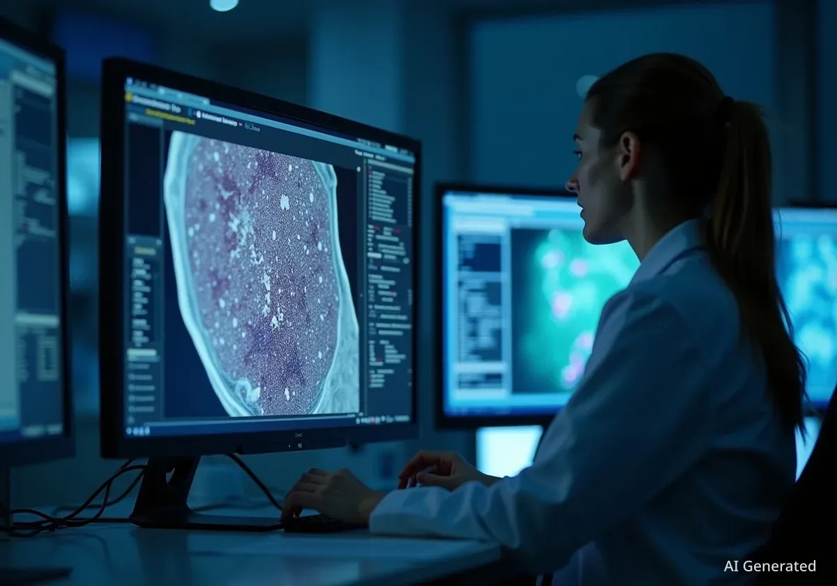


Researchers at the Massachusetts Institute of Technology (MIT) have created a new artificial intelligence system designed to significantly reduce the time required to analyze medical images. The tool, called MultiverSeg, allows clinical researchers to quickly and accurately highlight areas of interest in biomedical scans, a critical step for studying disease progression and evaluating new treatments.
Key Takeaways
- MIT researchers developed MultiverSeg, an AI tool that simplifies the annotation of medical images.
- The system learns from user interactions, progressively reducing the need for manual input.
- Unlike other models, it does not require users to have machine learning expertise or pre-segmented data for training.
- The tool aims to accelerate clinical studies, lower research costs, and improve efficiency in medical applications.
Addressing a Major Research Bottleneck
In medical research, one of the most fundamental tasks is image segmentation. This process involves outlining specific structures or regions within a medical scan, such as an organ or a tumor, to analyze changes over time.
For example, to study how a disease affects the brain's hippocampus, a scientist must manually trace its outline in numerous scans. This manual process is exceptionally time-consuming and can be a significant bottleneck in research, limiting the scope and speed of clinical studies.
What is Image Segmentation?
Image segmentation is the process of partitioning a digital image into multiple segments or sets of pixels. In medicine, this means identifying and outlining specific anatomical structures or abnormalities in scans like MRIs or X-rays. This detailed annotation is essential for quantitative analysis in research and diagnostics.
Existing methods for segmentation present challenges. Fully interactive tools require researchers to repeat the annotation process for every single image. Alternatively, creating a custom AI model requires manually segmenting hundreds of images for a training dataset, a complex process that demands significant time and machine-learning expertise.
An Interactive and Adaptive System
The new MIT system, MultiverSeg, offers a more efficient solution by combining the best features of existing approaches. It allows a researcher to annotate an image by providing simple inputs, such as clicking on points, drawing scribbles, or placing boxes around the area of interest.
The AI uses these interactions to generate a precise segmentation. The key innovation is that MultiverSeg remembers each annotated image, adding it to a "context set." When a new image is presented, the model uses this growing set of examples to make a better prediction with less user guidance.
As the researcher annotates more images, the system becomes progressively smarter. The number of required interactions diminishes until, eventually, the model can accurately segment new images with no user input at all.
"Many scientists might only have time to segment a few images per day for their research because manual image segmentation is so time-consuming. Our hope is that this system will enable new science by allowing clinical researchers to conduct studies they were prohibited from doing before because of the lack of an efficient tool," stated Hallee Wong, an MIT graduate student and lead author of the research paper.
This learning capability means users do not need to retrain the model for each new task. They can simply upload a new type of medical image and begin the interactive annotation process immediately, making the tool highly accessible and versatile.
Demonstrated Performance and Efficiency
In comparative tests, MultiverSeg outperformed other state-of-the-art tools for interactive image segmentation. The system's main advantage is its ability to reduce the user's workload over time.
Significant Reduction in Effort
According to the research, by the time a user annotated the ninth image in a new dataset, MultiverSeg required only two clicks to produce a segmentation more accurate than a model specifically designed for that task.
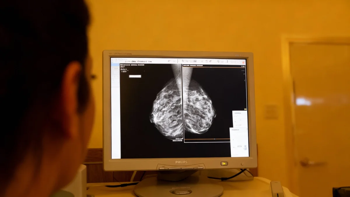
For certain types of images, such as X-rays, the researchers found that the model could achieve high accuracy on its own after the user manually segmented just one or two initial images. This represents a dramatic increase in efficiency.
Compared to a previous MIT-developed tool, ScribblePrompt, the new system achieved 90% accuracy with significantly less effort:
- It required roughly two-thirds the number of scribbles.
- It needed only three-fourths the number of clicks.
The interactive nature of the tool also allows users to correct any mistakes made by the AI. They can provide additional clicks or scribbles to refine the predicted outline until it meets the required level of precision. "With MultiverSeg, users can always provide more interactions to refine the AI predictions," Wong added. "This still dramatically accelerates the process because it is usually faster to correct something that exists than to start from scratch."
Potential Impact on Medicine and Research
By making image segmentation faster and more accessible, MultiverSeg could have a broad impact on medical science. It has the potential to accelerate studies on new treatments by allowing researchers to analyze larger datasets more quickly.
This efficiency could also help reduce the costs associated with clinical trials and other forms of medical research. Furthermore, the tool could be adapted for direct clinical use, assisting physicians in tasks such as planning radiation treatments, where precise outlining of tumors and healthy organs is critical.
The research team, which includes John Guttag and senior author Adrian Dalca, plans to test MultiverSeg in real-world scenarios with clinical partners. Their future work will focus on gathering user feedback to improve the system and extending its capabilities to handle 3D biomedical images, which are common in modern medical diagnostics.
The research is set to be presented at the International Conference on Computer Vision and was supported by organizations including the National Institutes of Health.